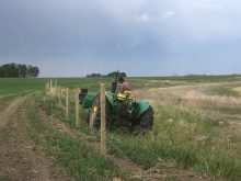

Most of the high water across the eastern Prairies will recede in the upcoming weeks, if they haven’t already. When cattle can be returned to these once-flooded pastures, the ground will probably be soaked, which will likely expand the territories of many cattle parasites such as the common deer liver fluke.
To combat this invasive species, beef producers should look for signs of liver flukes in their cattle, know the options for treating affected cattle and even employ some protocols to prevent it.
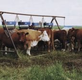
The funny thing is that many producers with infected cattle don’t even realize that their cattle have liver flukes. That’s because its symptoms are general in nature such as weight loss, reduced body condition, anemia and chronic diarrhea. Cattle are also susceptible to clostridial diseases such as blackleg and redwater, which can affect animals on their own and may not be associated with liver flukes.
Read Also
Get ready for an eventual transition on cattle traceability
When new federal cattle traceability rules do ultimately take effect, reporting requirements will vary for producers, transporters, feedlots and markets — but most of the onus will be on producers.
A friend that operates a 100-beef cow operation near the U.S. border told me that he had never seen a liver fluke before he butchered a young heifer that broke her leg.
When he eviscerated her and removed her liver; a few flat worms that were about three inches long, one inch wide and flat enough to see through tried to escape. With the help of his vet, they identified it as the deer liver fluke (fasciola magna).
Deer liver flukes live most of their lives in wet lowland regions, which include muddy areas and those areas with existing surface water often found in cattle pastures. All of which accommodate its life cycle from the time the fluke egg is initially shed in the manure of infected cows onto pasture until a new generation of eggs takes about 16 to 24 weeks.
Parasites have four stages
Subsequently, there are four major stages from fluke egg to hermaphroditic (contain both sex organs) adult liver flukes, which ultimately reside in the bile duct of cattle:
1. Egg stage: Eggs are shed in the manure of the beef cow from adult liver flukes.
2. Miracidium stage: Eggs hatch into free-swimming, ciliated (non-feeding) larvae called the miracidium. Within hours of hatching, the miracidium find and penetrate snails living on wet pastures.
3. Cercariae stage: Within the snail, one miracidium develops into hundreds of tadpole-like second larvae stage. After they leave their snail host, they attach to grass and become encysted metacercaria (later stage larva enclosed in a cyst).
4. Adult stage: The grazing cow ingests the encysted metacercaria contaminated grass. Once ingested, juvenile liver flukes are released from metacercaria capsules, penetrate the intestinal wall and burrow their way to and through the liver, which takes up to eight weeks. These fluke larvae enter the bile duct, where they mature into egg-laying adults to repeat the cycle.
Treatment options
Treatment comes down to a few flukicides: Albendazole (Valbazen), Corsulon (Ivomec Plus), and Triclabendazole (United Kingdom). The first two flukicides are only effective against mature adult liver flukes. Triclabendazole is the only product effective against very early immature flukes (two weeks old) through adulthood. There is no available flukicide treatment against shed eggs in cow manure.
For example, Albendazole is to be drenched for its first treatment for confirmed fluke infections in cattle in autumn. This is to remove any buildup of adult flukes that matured over the grazing season and when snails are dominant and less of a transmission threat. A second treatment is then given the following April/May, which reduces the number of egg-shedding adults before letting cattle out on pasture. It is noted that Albendazole should not be given to any pregnant cow or replacement heifer during the first 45 days of gestation, otherwise it may cause early embryonic deaths.
Again, identification of liver fluke infection is going to play a major role in its effective treatment. Rather than rely on egg counts, some British sheep and beef specialists have been collecting blood samples of suspect herds and testing for specific antibodies triggered by liver flukes. While the tests show promise in liver fluke identification, they point out that these antibodies only indicate cattle exposure, not active infection. There also might be interference by clostridial antibodies, which yields false positive results.
Regardless, this is the step in the right direction and valuable information. If one or two animals show a positive result, then the producer should work with their veterinarian to determine its validity and need for further testing. In this way, it should dovetail into a more effective treatment program against invading liver flukes on pasture.